VE-821 ATR Kinase Inhibition Suppresses Proliferation in Hypoxic Tumor Microenvironments
Copy code to add this image to your page
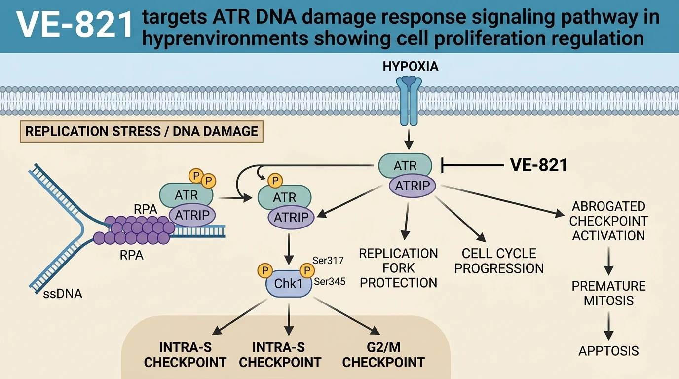
Solid tumor masses frequently develop severe pathophysiological microenvironments characterized by gradients of oxygen depletion, nutrient starvation, and acidic metabolite accumulation. Within these hypoxic microhabitats, tumor cells adapt their DNA damage response (DDR) pathways to maintain genomic stability and preserve proliferative capacity. Among the primary apical transducers of the DDR, Ataxia Telangiectasia and Rad3-related (ATR) kinase acts as the central sensor of replication stress and single-stranded DNA breaks. Understanding how ATR signaling functions within the physiological architecture of three-dimensional tissue remains critical for refining DDR-targeted oncology compounds.
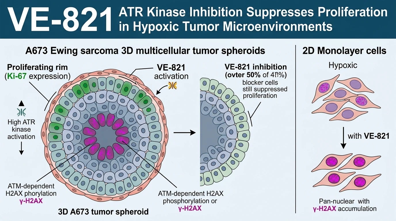
Traditional two-dimensional monolayer cultures fail to recapitulate the complex microenvironmental heterogeneity of solid tumors. To bridge this physiological gap, multicellular tumor spheroids (MCTS) provide a structured 3D model featuring outer proliferating rims, intermediate quiescent zones, and central hypoxic or necrotic cores. In A673 Ewing sarcoma spheroids, spatial mapping reveals a distinct functional division between apical DDR kinases under severe hypoxia. Ataxia Telangiectasia Mutated (ATM) kinase activity concentrates within the peri-necrotic core, directly driving the baseline phosphorylation of histone H2AX (γ-H2AX). Conversely, ATR kinase operates predominantly within the viable, outer regions of the spheroid mass, where it sustains ongoing DNA replication and cellular division.
Targeting ATR in this 3D model using the selective small-molecule inhibitor VE-821 provides clear evidence of this spatial specialization. Application of VE-821 to A673 spheroids led to a marked reduction in Ki-67-positive proliferating cells, exceeding a 50% decrease relative to vehicle controls. Spheroid growth kinetics were significantly retarded over extended culture periods. Remarkably, despite this profound anti-proliferative effect, pharmacological inhibition by VE-821 caused negligible changes in γ-H2AX signal intensity within the 3D spheroid core. This observation indicates that baseline H2AX phosphorylation in the central hypoxic domain remains reliant on ATM rather than ATR signaling.
A markedly different pharmacodynamic profile emerges when evaluating ATR inhibition in standard 2D monolayer cultures under equivalent hypoxic conditions. Treatment of 2D monolayer A673 cells with VE-821 induced robust pan-nuclear and focal γ-H2AX accumulation throughout the cell population. This striking divergence between 2D and 3D systems highlights the confounding effect of culture dimensionality on signaling readouts. In monolayer conditions, uniform oxygen deprivation combined with ATR inhibition causes acute replication fork collapse across all cells, yielding widespread H2AX phosphorylation. Within the 3D spheroid matrix, structural cell-cell interactions and physiological oxygen gradients buffer cells against uniform catastrophic fork collapse, restricting the phenotypic consequence of ATR inhibition primarily to proliferative arrest.
These empirical findings position ATR not as a uniform mediator of core tissue damage, but as a crucial engine of cell proliferation in hypoxic microenvironments. Evaluating ATR inhibitors like VE-821 within 3D spheroid models demonstrates that therapeutic responses are heavily influenced by spatial microarchitecture and localized kinase redundancy.
Spatial DDR Disconnect and Proliferative Arrest Dynamics
Copy code to add this image to your page
VE-821 selectively inhibits ATR kinase, disrupting replication stress responses under hypoxic conditions. In 3D Ewing sarcoma spheroids, ATR signaling primarily sustains cell proliferation, whereas ATM mediates H2AX phosphorylation in the necrotic core. Treatment with VE-821 reduces Ki-67-positive proliferating cells by over 50% and impairs spheroid expansion without altering peri-necrotic γ-H2AX levels. Conversely, in 2D hypoxic cultures, ATR inhibition induces widespread γ-H2AX expression. This mechanistic divergence emphasizes ATR as a specific driver of hypoxic cell division and illustrates the necessity of 3D cellular models for evaluating DDR-targeted cancer therapeutics.
ATR Inhibition Depletes Ki-67 Proliferative Fractions in Spheroids
Multicellular tumor spheroids recreate the spatial nutrient and oxygen gradients observed in avascular tumor regions. In A673 Ewing sarcoma spheroids, cell proliferation relies heavily on functional ATR kinase signaling. Pharmacological blockade using VE-821 suppressed Ki-67-positive proliferating fractions by more than 50% under experimental testing conditions. Growth kinetics of the 3D spheroid mass were markedly retarded over time. Interestingly, core phosphorylation of H2AX remained largely undisturbed within the 3D architectural matrix. ATM kinase activity independently maintains baseline H2AX phosphorylation in the peri-necrotic zone—a finding confirmed by targeted biomarker assays. Consequently, ATR operates predominantly as an engine of cellular replication rather than a sole mediator of baseline core DNA damage response within 3D hypoxic environments.
Divergent H2AX Phosphorylation Profiles Across 2D and 3D Microenvironments
Culturing A673 cells in 2D hypoxic monolayers yielded a distinctly different DDR phenotypical baseline. Administration of VE-821 induced intense pan-nuclear and focal γ-H2AX signal accumulation within 2D cultures. This contrast highlights how dimensionality alters DNA damage signaling cascades under oxygen deprivation. Monolayers lack the extracellular matrix architecture and tissue-level microenvironmental buffering present in 3D spheroids. Thus, spatial organization dictates cellular reliance on specific DDR nodes during hypoxic stress. Evaluating targeted ATR inhibitors in three-dimensional tissue models provides crucial predictive insights that standard two-dimensional assays fail to capture.