Bay K 8644 and Steroidal Analogues Driven L-Type Calcium Channel Activation in Ischemia
Copy code to add this image to your page
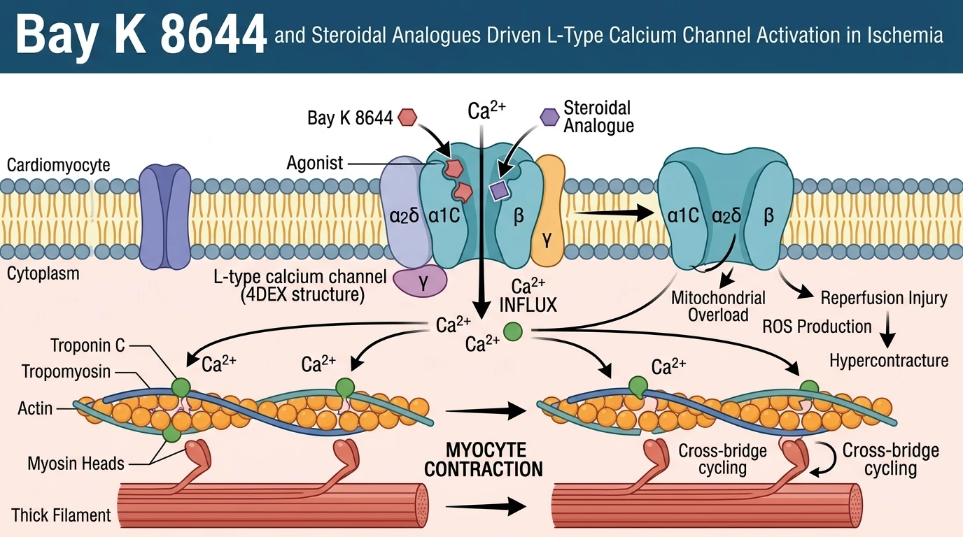
Myocardial ischemia/reperfusion injury represents a primary driver of mechanical failure and tissue loss following acute coronary occlusion. Restoring coronary blood flow paradoxically triggers intracellular ion dysregulation, mitochondrial membrane depolarization, and contractile dysfunction within cardiac myocytes. Functional recovery depends on maintaining controlled transsarcolemmal ion fluxes during the critical reperfusion window. L-type calcium channels (CaV1.2) govern the influx of extracellular Ca2+ required for excitation-contraction coupling. Small molecules that modulate these pore-forming proteins can alter channel open-state probability and dynamic ion conductance.
Pharmacological studies evaluating therapeutic compounds frequently employ isolated rat heart models to examine direct myocardial responses without systemic neural or vascular interference. In a representative ex vivo Langendorff preparation, a bis-steroid-methanocyclobuta-naphthalene-dione derivative (designated as compound 3) was tested against ischemia/reperfusion injury. Perfusion with compound 3 prior to global ischemia produced a dose-dependent reduction in myocardial tissue necrosis. Infarct areas were measured using triphenyltetrazolium chloride staining, showing significant reductions relative to untreated control hearts. Concurrently, left ventricular pressure (LVP) increased in a concentration-dependent manner, confirming preserved contractile force during reperfusion.
The functional profile of compound 3 mirrors the pharmacodynamic signature of dihydropyridine receptor agonists. The reference agonist Bay K 8644 serves as the standard benchmark for positive inotropic activity via dihydropyridine site activation. In parallel isolated heart perfusions, compound 3 produced positive inotropic responses equivalent in magnitude to those generated by Bay K 8644. To establish whether these contractile gains were mediated strictly through dihydropyridine receptors, hearts were co-treated with the L-type calcium channel blocker nifedipine. Nifedipine administration completely abolished the positive inotropic effects and LVP elevation induced by compound 3. This total competitive antagonism demonstrates that the cardioprotective actions rely on direct activation of the dihydropyridine binding locus rather than secondary adrenergic signaling.
To understand the atomic basis of this activation, computational modeling was conducted using crystal structures of the channel protein. Structural coordinates from the L-type calcium channel model (PDB ID: 4DEX) provided the structural template for ligand docking simulations. Compound 3 adopted a low-energy conformation within the transmembrane pore domain. Docking calculations revealed key electrostatic contacts and hydrophobic interactions with residue side chains forming the dihydropyridine binding pocket. These contact points overlap substantially with the binding trajectory of Bay K 8644. Ligand binding at this site stabilizes the channel pore in an open conformation, facilitating controlled Ca2+ entry. Integrating these atomic insights into broader downstream networks, such as the core calcium signaling pathway architecture, clarifies how structural modifications on a bis-steroid backbone retain receptor affinity while preserving ventricular mechanical stability during ischemic stress.
Pharmacological Agonism and Myocardial Hemodynamic Responses
Copy code to add this image to your page
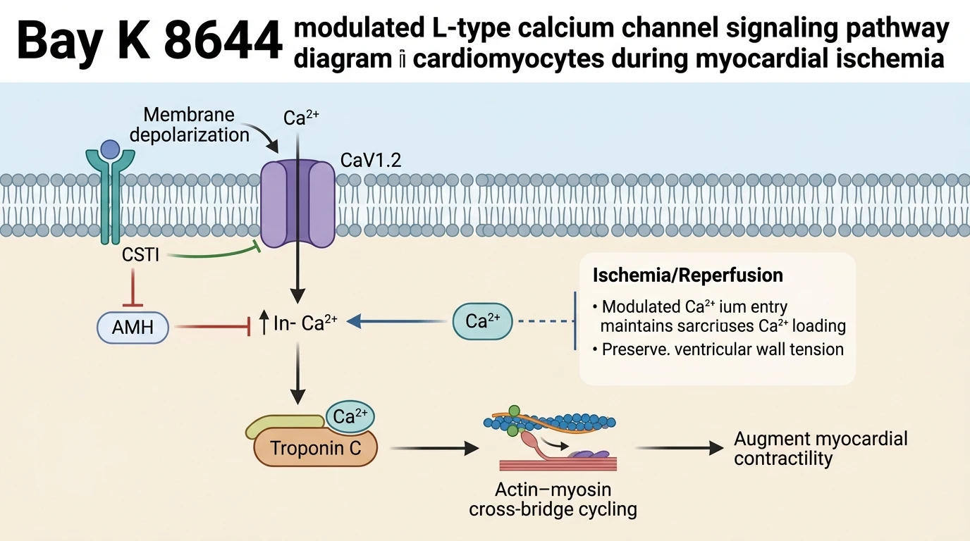
Agonism of L-type calcium channels by Bay K 8644 and structural analogues promotes transsarcolemmal Ca2+ entry into cardiac myocytes. Binding within the pore domain of the CaV1.2 alpha-1 subunit stabilizes the open state, restoring left ventricular pressure and reducing tissue necrosis after ischemic insult.
Dose-Dependent Inotropic Recovery in Isolated Hearts
Hemodynamic recovery after ischemic arrest depends on immediate calcium availability within the sarcoplasm. In ex vivo rat heart perfusions, administration of compound 3 restored developed pressure across multiple coronary flow rates. Left ventricular pressure rose progressively with increasing compound concentrations. Peak pressure gains matched those induced by Bay K 8644 at equivalent micromolar doses. Infarct size diminished proportionally. Tissue viability assays confirmed preserved myocardial architecture within the left ventricular wall. Under these ex vivo conditions, positive inotropy occurred without accelerating resting diastolic tone.
Nifedipine Blockade Confirms Target Channel Specificity
Receptor specificity was evaluated by introducing competitive channel antagonists into the perfusion buffer. Co-administration of nifedipine fully suppressed the inotropic response of compound 3. Contractile force returned to baseline levels immediately following antagonist infusion. The absolute reversal by nifedipine verifies that signaling occurs strictly through CaV1.2 subunits. Docking analysis into the 4DEX channel structure corroborated these functional findings. Residue contacts within the pore domain aligned with the binding mode of Bay K 8644. These shared steric boundaries confirm a common dihydropyridine-site mechanism.