S63845 Selective MCL-1 Inhibition Restores Intrinsic Apoptosis in Venetoclax-Resistant Malignancies
Copy code to add this image to your page
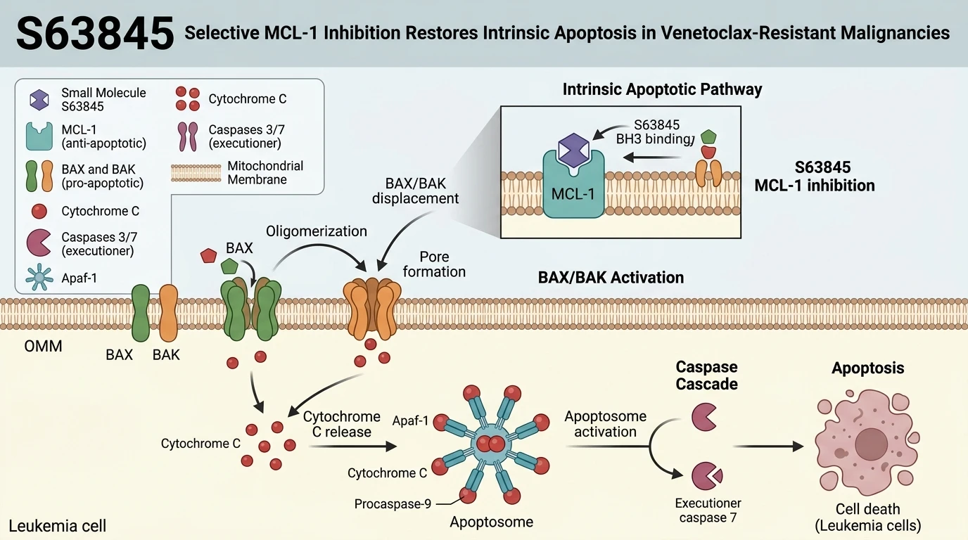
Targeted inhibition of anti-apoptotic machinery represents a key strategy in overcoming chemotherapy evasion in hematologic malignancies. Myeloid cell leukemia-1 (MCL-1), a structural member of the BCL-2 anti-apoptotic protein family, acts as a critical survival factor by binding and sequestering pro-apoptotic proteins such as BIM, NOXA, and the pore-forming effector BAK. In disease models including acute myeloid leukemia (AML) and myelodysplastic syndromes (MDS), high MCL-1 expression directly counteracts single-agent therapies. Most notably, selective BCL-2 inhibition via venetoclax frequently induces operational reliance on MCL-1, creating an axis of acquired resistance that prevents outer mitochondrial membrane permeabilization.
S63845 was identified as a potent, synthetic small-molecule inhibitor engineered to target the BH3-binding groove of MCL-1 with sub-nanomolar affinity (K_d < 1 nM). Structural co-crystallography demonstrates that S63845 occupies the hydrophobic P2 and P4 pockets of MCL-1, inducing conformational shifts that selectively disrupt binding to endogenous BH3 ligands. Unlike non-selective BCL-2 family antagonists, S63845 exhibits multi-log selectivity for MCL-1 over closely related homologs BCL-2 and BCL-xL under tested in vitro binding conditions. This targeted displacement liberates effector proteins BAX and BAK from sequestration.
At the cellular level, engagement of MCL-1 by S63845 initiates the classical intrinsic apoptotic cascade. Free BAK and BAX undergo conformational activation and homooligomerization, assembling into high-molecular-weight proteolipid pores within the mitochondrial outer membrane. This pore formation drives mitochondrial outer membrane permeabilization (MOMP), causing irreversible loss of mitochondrial transmembrane potential. Soluble apoptogenic factors, including cytochrome c and SMAC/DIABLO, flux from the intermembrane space into the cytosol. Cytosolic cytochrome c complexes with APAF-1 and dATP to form the heptameric apoptosome, recruiting and activating procaspase-9. Active CASP9 subsequently cleaves downstream executioner caspases CASP3 and CASP7, resulting in systematic cleavage of structural substrates such as poly(ADP-ribose) polymerase (PARP-1) and eventual cell demise.
In preclinical primary AML blasts and cell line screens, administration of S63845 consistently triggered apoptosis across diverse genetic backgrounds, including FLT3-ITD and TP53-mutated contexts. Experimental models demonstrate that combined inhibition of BCL-2 and MCL-1 achieves synergistic cytotoxicity in venetoclax-resistant cell lines by neutralizing redundant survival mechanisms. However, therapeutic sensitivity correlates strictly with intracellular MCL-1 dependency ratios; primary samples with high relative BCL-xL expression exhibit reduced susceptibility unless co-inhibited. These findings define the primary pharmacodynamic identity of S63845 as a targeted disinhibitor of the intrinsic apoptotic apparatus.
Pharmacological Characterization and Apoptotic Resensitization Dynamics
Copy code to add this image to your page
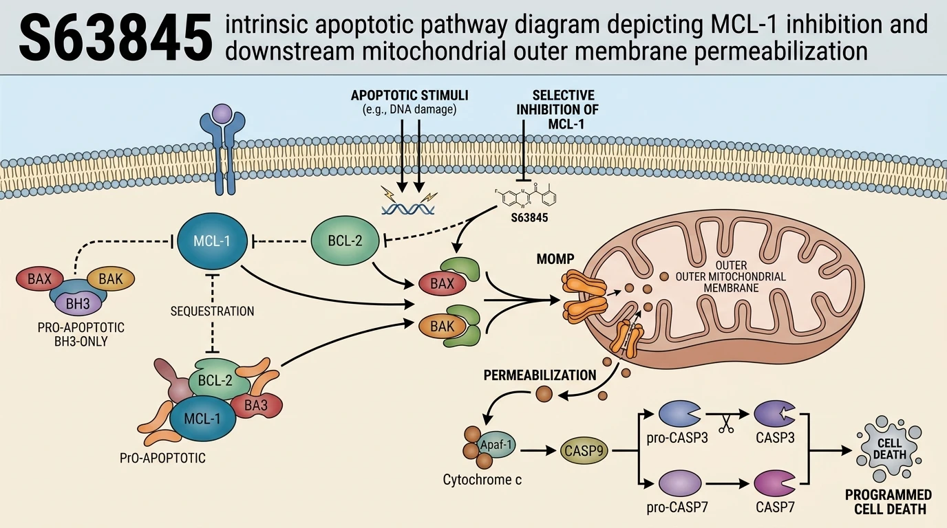
S63845 is a selective small-molecule inhibitor targeting the anti-apoptotic protein MCL-1 with sub-nanomolar affinity. By occupying the BH3-binding groove of MCL-1, S63845 displaces pro-apoptotic proteins such as BIM, BAX, and BAK. The released pro-apoptotic effectors form oligomers on the outer mitochondrial membrane, causing mitochondrial outer membrane permeabilization (MOMP). This leads to cytochrome c release, caspase cascade activation, and intrinsic apoptosis. Consequently, S63845 overcomes MCL-1-mediated resistance to selective BCL-2 inhibitors like venetoclax in hematologic malignancies such as acute myeloid leukemia and myelodysplastic syndromes.
High-Affinity Engagement Neutralizes MCL-1 in AML Blasts
Disruption of anti-apoptotic node interactions remains central to targeting hematologic malignancies. S63845 binds directly to the canonical hydrophobic BH3-binding groove of MCL-1 with sub-nanomolar affinity. This high-affinity interaction displaces endogenous pro-apoptotic BH3-only proteins—specifically BIM and NOXA—from the inhibitor-binding pocket. Inhibitory complex dissociation occurs rapidly in patient-derived AML cell lines. Downstream effector sequestration ceased within hours. Consequently, free pro-apoptotic factors migrate to the mitochondrial outer membrane. While these binding kinetics demonstrate high specificity in vitro, target occupancy thresholds in heterogeneous solid tumor microenvironments remain to be fully characterized.
Bifunctional BAX/BAK Activation Overcomes Venetoclax Resistance Models
Resistance to monotherapy BCL-2 inhibition by venetoclax frequently arises through compensatory upregulation of MCL-1. In myelodysplastic syndrome models displaying high baseline MCL-1 expression, venetoclax fails to induce complete MOMP. Co-treatment or sequential administration of S63845 effectively neutralizes this compensatory survival axis. Unbound BAX and BAK undergo conformational rearrangement and homooligomerization. Mitochondrial membrane potential collapsed rapidly across evaluated primary blast cohorts. Cytochrome c release triggered robust cleavage of procaspase-9 and procaspase-3. Cellular viability dropped sharply following combination therapy in primary patient isolates. Notably, these synthetic lethal interactions depend on baseline expression ratios of BCL-xL and MCL-1 within specific lineage subtypes.
Pharmacodynamic Biomarkers Confirm On-Target Apoptotic Cascades in Vivo
In murine xenograft models harboring refractory AML cells, administration of S63845 demonstrated clear target engagement without overt systemic toxicity. Cleaved PARP elevation served as a primary pharmacodynamic marker of ongoing apoptotic execution. Tumor burden decreased substantially within seven days of treatment initiating. The therapeutic window remained intact across tested dosing schedules. Phosphorylation of downstream survival signaling nodes remained unaltered, confirming target selectivity against off-target kinases. Observations in these preclinical models suggest that durable responses require sustained target saturation. Further translational studies are necessary to evaluate these responses in randomized clinical cohorts.