AICAR (Acadesine) Activates AMPK Signaling to Reverse Cachectic Muscle Wasting
Copy code to add this image to your page
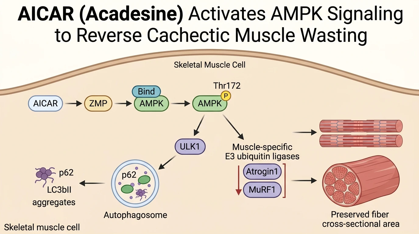
Cancer-associated cachexia manifests as a debilitating metabolic syndrome characterized by progressive loss of skeletal muscle mass, systemic inflammation, and severe functional impairment. In the C26 colon carcinoma murine model, cachectic muscle wasting driven by tumor-derived inflammatory cytokines exhibits profound alterations in protein turnover kinetics. Rather than resulting solely from elevated proteolysis, recent evidence demonstrates that severe muscle wasting in C26-bearing mice stems from a blockage in autophagic flux. Autophagy represents the primary catabolic mechanism for clearing damaged organelles and macromolecular complexes. Under basal physiological conditions, autophagosome formation and lysosomal clearance remain tightly balanced. In cachectic gastrocnemius muscle, however, biochemical profiling demonstrates a marked elevation of lipidated LC3bII alongside p62/SQSTM1 protein accumulation. This concurrent accumulation indicates an interruption in late-stage autophagic clearance, leading to toxic intracellular protein aggregation and muscular atrophy.
To exacerbate this metabolic destabilization, cachectic muscle tissues display pronounced transcriptional upregulation of muscle-specific E3 ubiquitin ligases, specifically Atrogin1 (FBXO32) and MuRF1 (TRIM63). These ubiquitin ligases selectively target structural myofibrillar proteins, including myosin heavy chain and actin, for 26S proteasomal degradation. Pharmacological activation of AMP-activated protein kinase (AMPK) using AICAR (Acadesine) mimics the biochemical strain of voluntary endurance exercise. AICAR enters skeletal muscle cells via nucleoside transporters, where adenosine kinase phosphorylates it into 5-aminoimidazole-4-carboxamide ribonucleotide (ZMP). As an AMP analog, ZMP binds directly to the gamma subunit of AMPK, inducing a conformational change that promotes Thr172 phosphorylation by upstream kinases such as LKB1.
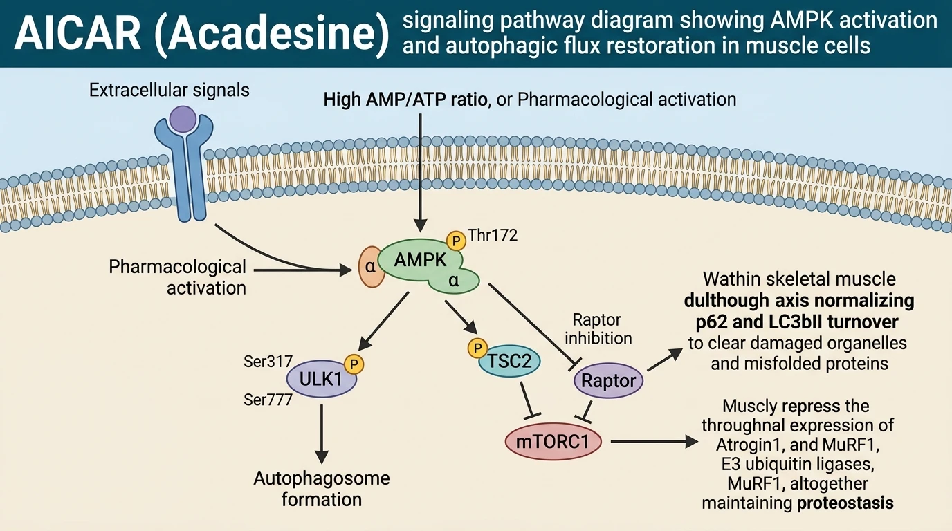
Once activated, AMPK exerts dual control over the autophagic machinery. First, AMPK directly phosphorylates the pro-autophagic initiator kinase ULK1 at Ser317 and Ser777, activating the inductive phase of autophagosome biogenesis. Second, AMPK antagonizes mechanistic target of rapamycin complex 1 (mTORC1) activity via phosphorylation of tuberous sclerosis complex 2 (TSC2) at Ser1387 and direct phosphorylation of Raptor. By relieving mTORC1-mediated inhibition of ULK1, AMPK re-establishes vesicular movement and lysosomal fusion. Daily administration of AICAR to C26 carcinoma-bearing mice reactivates stalled autophagic flux, promoting the rapid clearance of p62 and normalizing LC3bII/LC3bI ratios within skeletal muscle tissue.
Critically, the restoration of autophagic flux by AICAR directly impacts the expression of E3 ubiquitin ligases. Re-establishing vesicular clearance suppresses the hyperactivation of Atrogin1 and MuRF1 transcripts, halting accelerated myofibrillar breakdown. Histological analysis of tibialis anterior and gastrocnemius muscles from AICAR-treated cachectic mice reveals significant preservation of skeletal muscle wet weight and myofiber cross-sectional area (CSA) relative to vehicle-treated controls. In vitro studies using C2C12 myotubes exposed to cachectic tumor-conditioned media validate these systemic findings; treatment with AICAR prevents myotube diameter shrinkage and stabilizes intracellular protein content. Similar outcomes observed with rapamycin confirm that restoring autophagic turnover represents a central node in preserving muscle architecture during tumor progression. Pharmacological target engagement of AMPK by AICAR successfully replicates the protective metabolic adaptations of physical exercise, offering a concrete biochemical strategy to counteract catabolic muscle loss in cancer cachexia.
Pharmacological AMPK Activation and Myofibrillar Proteostasis Maintenance
Copy code to add this image to your page
AICAR functions as an exercise mimetic that directly activates AMP-activated protein kinase (AMPK) via intracellular conversion to ZMP. In C26 carcinoma-induced cachexia models, AMPK activation re-establishes blocked autophagic flux characterized by excessive LC3bII and p62 protein accumulation. By reinstating autophagosome clearance and repressing the muscle-specific E3 ubiquitin ligases Atrogin1 and MuRF1, AMPK activation reduces excessive myofibrillar protein breakdown. Consequently, skeletal muscle mass and muscle fiber cross-sectional area are preserved, demonstrating that pharmacological stimulation of autophagic turnover effectively counteracts cancer-associated muscle wasting.
Atrogene Downregulation and Autophagic Clearance in C26 Carcinoma Models
In murine C26 colon carcinoma models, severe muscle cachexia correlates with disrupted autophagic degradation pathways. Quantitative western blot analyses demonstrate marked accumulation of both p62/SQSTM1 and lipidated LC3bII within gastrocnemius muscle lysates. This dual elevation signifies impaired autophagic flux rather than pure pathway inhibition. Daily administration of Acadesine (100 mg/kg) restored lysosomal clearance kinetics. Phosphorylation of AMPK at Thr172 increased sharply following treatment; this biochemical change prompted downstream ULK1 phosphorylation at Ser317. Consequently, p62 levels declined to baseline parameters—indicating complete turnover of autophagic vesicles. Furthermore, transcripts for the muscle-specific E3 ubiquitin ligases Atrogin1 and MuRF1 decreased significantly following pharmacological AMPK engagement. The suppression of these key atrogenes reduced targeted degradation of structural sarcomeric proteins such as myosin heavy chain.
Preservation of Myofiber Cross-Sectional Area Under Hypercatabolic Conditions
Morphometric evaluation of skeletal muscle tissue revealed substantial protection against cancer-induced atrophy. Histological staining of tibialis anterior cross-sections demonstrated that untreated C26 tumor-bearing mice suffered an approximate 35% reduction in mean myofiber cross-sectional area. In contrast, systemic administration of Acadesine effectively preserved fiber dimensions. Muscle fiber diameter remained within normal physiological ranges despite high systemic tumor burden; this structural preservation directly correlated with enhanced force generation capacity in ex vivo muscle preparations. In parallel in vitro studies utilizing C2C12 myotubes exposed to cachectic conditioning media, treatment with Acadesine prevented myotube shrinkage without inducing overt cytotoxicity. These observations suggest that restoring autophagic flux through pharmacological AMPK activation mimics the muscular adaptations of voluntary exercise, establishing a viable mechanism to mitigate metabolic dysregulation in cancer cachexia.