Berzosertib (VE-822) Berzosertib ATM/ATR Kinase Inhibition Attenuates Lupus B Cell Hyperactivity
Copy code to add this image to your page
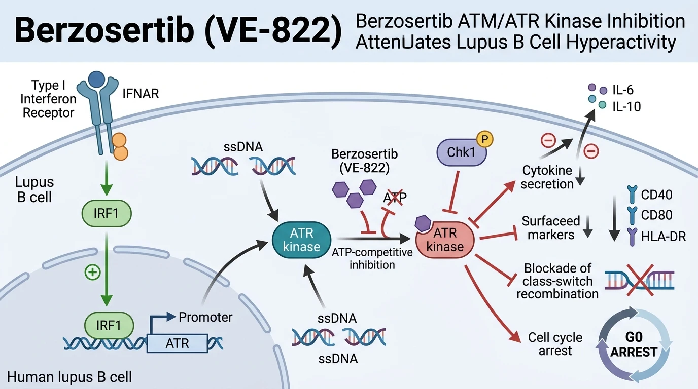
Systemic lupus erythematosus (SLE) is characterized by loss of self-tolerance and aberrant B cell activation, culminating in autoantibody production and multi-organ tissue damage. Among the central drivers of this pathology is an elevated type I interferon (IFN-α) gene signature. While IFN-α is known to modulate innate immune signaling, its direct convergence with genomic maintenance machinery in lymphocytes reveals a key regulatory axis. Recent experimental work identifies heightened activity of the ATR/Chk1 DNA damage response (DDR) pathway in primary peripheral B cells isolated from patients with active SLE. Elevated baseline DNA single-strand breaks and replication stress in auto-reactive lymphocytes recruit Ataxia telangiectasia and Rad3-related protein kinase (ATR) to damaged chromatin sites, where it orchestrates checkpoint activation.
Mechanistic investigation demonstrates that elevated IFN-α directly enhances ATR transcript and protein levels in primary B cells. Transcriptional profiling combined with promoter binding analysis reveals that interferon regulatory factor 1 (IRF1), induced downstream of type I interferon receptor engagement, binds to specific consensus motifs within the human ATR gene promoter. This transcriptional upregulation amplifies baseline DDR signaling, leading to constitutive phosphorylation of downstream effector Chk1 at Ser317 and Ser345. Rather than inducing apoptotic elimination, this persistent DDR state in SLE B cells promotes cellular survival, hyper-reactivity, and differentiation. To investigate whether this baseline axis can be pharmacologically reversed, researchers evaluated the potent and selective small-molecule ATR inhibitor Berzosertib (VE-822).
Selective small-molecule inhibition using Berzosertib (VE-822) effectively uncouples the IRF1-driven DDR cascade in patient-derived B cells under in vitro assay conditions. By competing with ATP at the ATR catalytic core, Berzosertib (VE-822) blocks Chk1 phosphorylation without interfering with parallel ATM kinase functions. Interruption of kinase domain structural signaling suppresses the pathological hyper-immunogenicity of SLE B cells. Specifically, ATR inhibition results in a marked decline in the secretion of key inflammatory and regulatory cytokines, including interleukin-6 (IL-6), interleukin-10 (IL-10), interleukin-4 (IL-4), and tumor necrosis factor-beta (TNF-β). Beyond cytokine suppression, pharmacological target engagement downregulates critical cell surface costimulatory markers and antigen-presentation machinery, notably CD40, CD80, and HLA-DR.
Functional downstream consequences extend to antibody synthesis and cell fate commitment. Treatment with Berzosertib (VE-822) severely impairs immunoglobulin class-switch recombination (CSR) and total IgG/IgM synthesis in stimulated patient lymphocytes. Furthermore, the differentiation of activated naive and memory B cells into CD19+CD27brightCD38bright plasmablasts is substantially reduced. Flow cytometric cell cycle analysis indicates that ATR blockade does not primarily trigger acute necrotic cell death; instead, it enforces a stable G0 resting cell cycle arrest. These empirical observations position the IRF1-ATR axis as a crucial molecular driver of pathogenic B cell hyper-reactivity, validating small-molecule ATR inhibitors like Berzosertib (VE-822) as candidate targeted immunomodulators for autoimmune conditions.
Targeting the IRF1-ATR DDR Axis in Autoimmune Lymphocytes
Copy code to add this image to your page
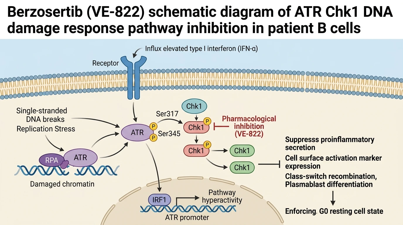
Systemic lupus erythematosus B cells exhibit hyperactivation of the ATR/Chk1 DNA damage response pathway driven by type I interferon. IFN-α signals through IRF1 to bind the ATR promoter, increasing ATR kinase expression. Berzosertib (VE-822) selectively inhibits ATR kinase activity, preventing downstream Chk1 phosphorylation. This pharmacological blockade downregulates proinflammatory cytokines (IL-6, IL-10, IL-4, TNF-β) and surface markers (CD40, CD80, HLA-DR), suppresses immunoglobulin class-switch recombination, and halts plasmablast differentiation by arresting cells in a G0 resting state.
Type I Interferon Drives Transcriptional Induction of ATR via IRF1
In primary B cells isolated from patients with active SLE, elevated type I interferon levels establish a chronic pro-inflammatory environment. Quantitative gene expression profiling shows a significant correlation between IFN-α expression and elevated ATR mRNA transcript levels. Chromatin immunoprecipitation assays demonstrate that interferon regulatory factor 1 (IRF1) directly occupies the proximal ATR promoter region. This binding increases ATR protein abundance, sensitizing B cells to endogenous replication stress. Application of the selective inhibitor in Berzosertib ATM/ATR intervention paradigms inhibits this kinase node directly. Cellular assays confirm that blocking ATR enzymatic activity disrupts downstream Chk1 Ser317 phosphorylation within two hours of exposure. Consequently, the hyper-activated DDR state induced by IFN-α is rapidly neutralized.
Pharmacological ATR Inhibition Enforces G0 Quiescence and Blocks Plasmablast Differentiation
Downstream of ATR activation, proliferating autoimmune B cells undergo rapid differentiation into antibody-secreting plasma cells. In cell culture models using patient-derived B cells stimulated with TLR7/9 agonists, treatment with Berzosertib (VE-822) profoundly shifts cell cycle dynamics. Proliferation assays reveal a complete arrest of stimulated lymphocytes in the G0/G1 resting phase. S-phase entry dropped significantly in treated samples. Utilizing Berzosertib ATM/ATR target engagement strategies effectively suppresses activation-induced cytidine deaminase (AID) expression, thereby blocking immunoglobulin class-switch recombination. Plasmablast generation was reduced by over 60 percent compared to vehicle controls, preventing the accumulation of short-lived autoantibody-secreting cells.
Attenuation of Proinflammatory Cytokines and Costimulatory Marker Expression
The pathogenic capacity of SLE B cells relies heavily on antigen presentation and paracrine signaling to helper T cells. Flow cytometry reveals that pharmacological ATR blockade leads to marked surface reduction of CD40, CD80, and HLA-DR molecules on primary B cells. Costimulatory capacity was markedly impaired. Enzyme-linked immunosorbent assays (ELISA) of culture supernatants demonstrate concurrent, dose-dependent reductions in secreted IL-6, IL-10, IL-4, and TNF-β levels. Importantly, viability assays indicate that these immunomodulatory effects occur without inducing unspecific cytotoxicity in non-activated control lymphocytes. These findings demonstrate that the IRF1-ATR pathway acts as an upstream metabolic and signaling checkpoint regulating autoimmune B cell effector functions.