Q-VD-Oph Suppresses Ischemia-Induced Neural Stem Cell Proliferation via Caspase Inhibition
Copy code to add this image to your page
Ischemic stroke remains a leading cause of long-term neurological disability worldwide. Acute arterial occlusion precipitates localized hypoxia, metabolic collapse, and progressive cell death within the affected cerebral architecture. Following focal ischemic injury, endogenous neurogenesis is transiently activated within adult neurogenic niches, most notably the subventricular zone (SVZ) adjacent to the lateral ventricles. Neural stem/progenitor cells (NSPCs) undergo proliferative expansion, generate committed neuroblasts, and migrate toward the peri-infarct cortex along chemokine gradients. However, the vast majority of newly generated neuroblasts fail to survive the harsh, cytotoxic post-stroke microenvironment, preventing substantial spontaneous functional recovery. Consequently, therapeutic strategies aiming to promote neural repair have frequently focused on suppressing apoptotic cell death mechanisms to enhance NSPC survival and neurogenesis.
Broad-spectrum caspase inhibition has represented a primary strategy for blocking programmed cell death post-ischemia. Among available pharmacological agents, Q-VD-Oph (quinolyl-valyl-aspartyl-difluorophenoxymethylketone) stands out as a highly potent, cell-permeable, and non-toxic pan-caspase inhibitor. Unlike early peptide-based inhibitors, Q-VD-Oph exhibits enhanced biochemical stability and reduced off-target toxicity, efficiently neutralizing effector caspases (caspase-3, -6, and -7) as well as initiator and inflammatory caspases (caspase-1, -8, and -9). Based on the classic paradigm that caspase activation triggers neuroblast apoptosis, systemic administration of Q-VD-Oph was hypothesized to protect vulnerable NSPCs, prolong neuroblast survival, and ultimately increase the population of mature, functional cortical neurons following ischemic insult.
Experimental evaluation in a murine model of focal cortical ischemia yielded unexpected results that challenge conventional assumptions regarding caspase blockade in neural tissue repair. Systemic post-ischemic treatment with Q-VD-Oph failed to induce any increase in the density of newborn mature cortical neurons. Quantitative histological and birth-dating label analyses demonstrated that neurogenesis within the injured cortical parenchyma remained unimproved despite robust systemic inhibition of caspase activity. This clear divergence between anti-apoptotic protection and cortical neuroblast accumulation prompted a detailed re-examination of how Q-VD-Oph affects the upstream activation and recruitment of NSPCs in vivo.
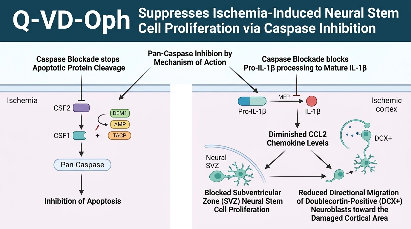
Cellular tracking within the neurogenic microenvironment demonstrated that Q-VD-Oph profoundly altered early post-stroke NSPC dynamics. In vehicle-treated ischemic controls, focal cortical infarction induced a robust proliferative surge of NSPCs within the SVZ, accompanied by the stream-like migration of doublecortin-positive neuroblasts toward the cortical lesion. In contrast, treatment with Q-VD-Oph completely abolished this ischemia-induced increase in SVZ NSPC proliferation. Furthermore, the total density of migratory neuroblasts reaching the damaged cortical tissue was severely depleted in Q-VD-Oph-treated animals. These observations demonstrate that caspase activity is not merely an executioner of cell death, but serves an indispensable non-apoptotic function in driving injury-induced NSPC expansion and directional recruitment.
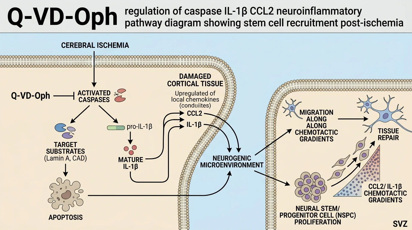
Mechanistic analysis revealed that Q-VD-Oph suppresses essential pro-inflammatory signaling nodes within the neurogenic microenvironment. Ischemic brain tissue typically produces a transient rise in interleukin-1β (IL-1β) and monocyte chemoattractant protein-1 (CCL2). Administration of Q-VD-Oph markedly attenuated cortical expression of both IL-1β and CCL2. Inflammatory caspases, specifically caspase-1, process inactive pro-IL-1β into its mature, biologically active cytokine form. Active IL-1β subsequently acts on stromal and glial elements to induce local CCL2 production, generating a chemoattractant gradient that drives NSPC division and neuroblast homing. By interrupting this cleavage-dependent cascade, Q-VD-Oph deprives NSPCs of critical inflammatory signals required for post-ischemic repair, underscoring the complex, dual nature of caspase signaling in acute neural injury.
Caspase Activity Regulates Post-Ischemic Neurogenic Niche Signals
Copy code to add this image to your page
Q-VD-Oph is a broad-spectrum, irreversible pan-caspase inhibitor that potently blocks apoptotic and inflammatory caspase catalytic domains. By inhibiting caspase catalytic activity, Q-VD-Oph prevents the cleavage-dependent maturation of pro-inflammatory cytokines such as IL-1β and suppresses downstream upregulation of the chemokine CCL2. In cortical ischemia models, this suppression disrupts the local neuroinflammatory signaling cascade required to trigger subventricular zone neural stem/progenitor cell proliferation and directional neuroblast migration to the site of injury.
Suppression of SVZ Proliferation and Neuroblast Migration
In ischemic mouse models, focal cortical infarction triggers a rapid surge in subventricular zone cell division. This compensatory proliferation fuels the production of migratory neuroblasts. Systemic administration of Q-VD-Oph completely abolished this injury-induced proliferative response within the subventricular zone. Quantitative doublecortin immunostaining confirmed a significant reduction in neuroblasts traversing the rostral migratory route toward the damaged cortex. Within this specific cellular context, pan-caspase inhibition did not improve neural stem cell survival. Instead, Q-VD-Oph impaired early mitotic recruitment. Cellular proliferation metrics dropped below sham levels in treated cohorts. These findings indicate that basal caspase activity is necessary to mobilize progenitor pools following acute brain injury.
Attenuation of Cortical CCL2 and Interleukin-1β Expressions
Neuroinflammatory signaling coordinates stem cell recruitment post-injury. Focal stroke elevated cortical concentrations of mature interleukin-1β and the chemoattractant chemokine CCL2 in vehicle controls. Treatment with Q-VD-Oph substantially decreased cortical levels of both signaling factors under tested in vivo conditions. Caspase-1 cleavage is required to convert pro-interleukin-1β into its active extracellular cytokine form. Active interleukin-1β subsequently stimulates astrocytic and microglial release of CCL2. By disrupting this processing cascade, Q-VD-Oph effectively flattened the chemoattractant gradient necessary to guide neuroblasts toward cortical lesions. Apoptosis inhibition alone proved insufficient to compensate for the loss of these crucial inflammatory neurogenic cues.