Oligomycin A (MCH 32) Oligomycin A Antineoplastic and I inhibitor F1Fo ATP Synthase Modulation
Copy code to add this image to your page
Mitochondrial bioenergetics rely fundamentally on the electrochemical proton gradient maintained across the inner mitochondrial membrane. The multiprotein F1Fo ATP synthase complex functions as the terminal enzyme of oxidative phosphorylation, harnessing this proton motive force to catalyze the synthesis of ATP from ADP and inorganic phosphate. Within this rotary macromolecular machine, the hydrophobic Fo sector mediates proton translocation across the lipid bilayer. Specifically, the ring composed of subunit c forms the essential conduit through which protons traverse. Targeted pharmacological disruption of this proton transport channel yields profound cellular bioenergetic consequences, altering cellular survival signaling and structural architecture.
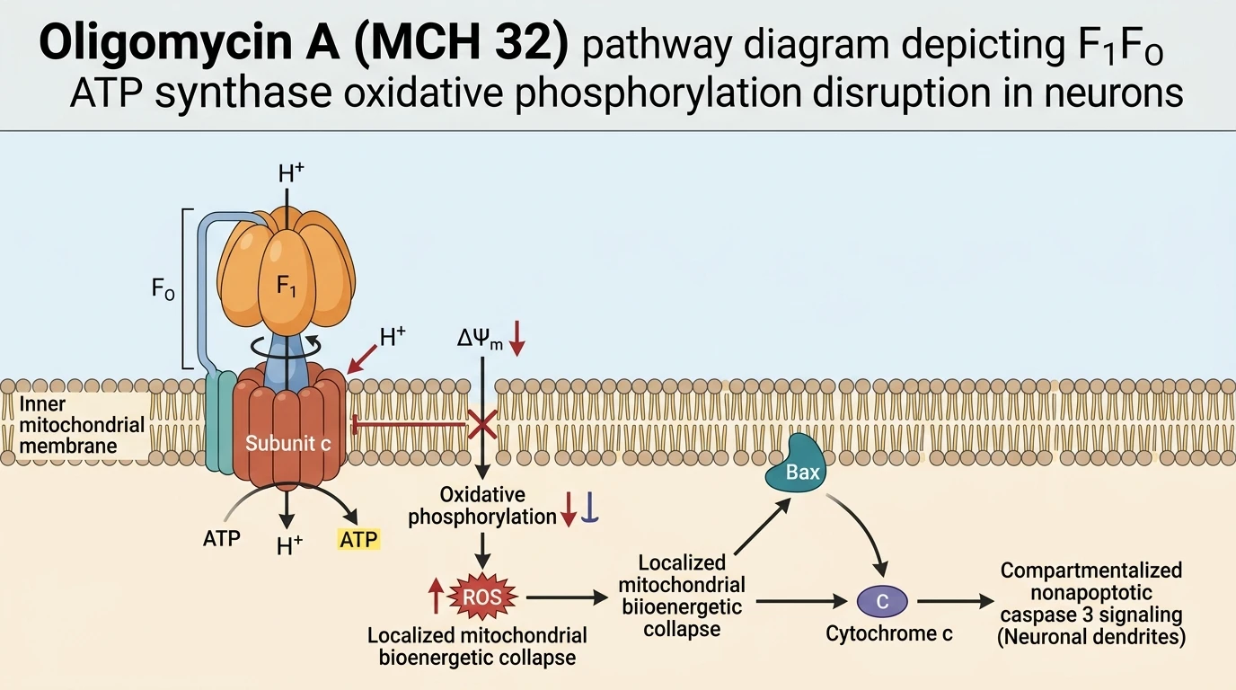
Oligomycin A (MCH 32) operates as a high-affinity, highly selective small-molecule inhibitor of F1Fo ATP synthase. By binding directly to subunit c within the Fo domain, the compound physically blocks proton conductance, uncoupling electron transport chain activity from ATP production. This specific target engagement renders the Oligomycin A Antineoplastic and I inhibitor profile a critical tool for dissecting the immediate downstream pathophysiological outcomes of acute bioenergetic failure. At submicromolar to micromolar concentrations, F1Fo ATP synthase (c subunit) inhibition does not merely induce generalized metabolic paralysis; rather, it initiates a spatially restricted signaling cascade within vulnerable cellular compartments.
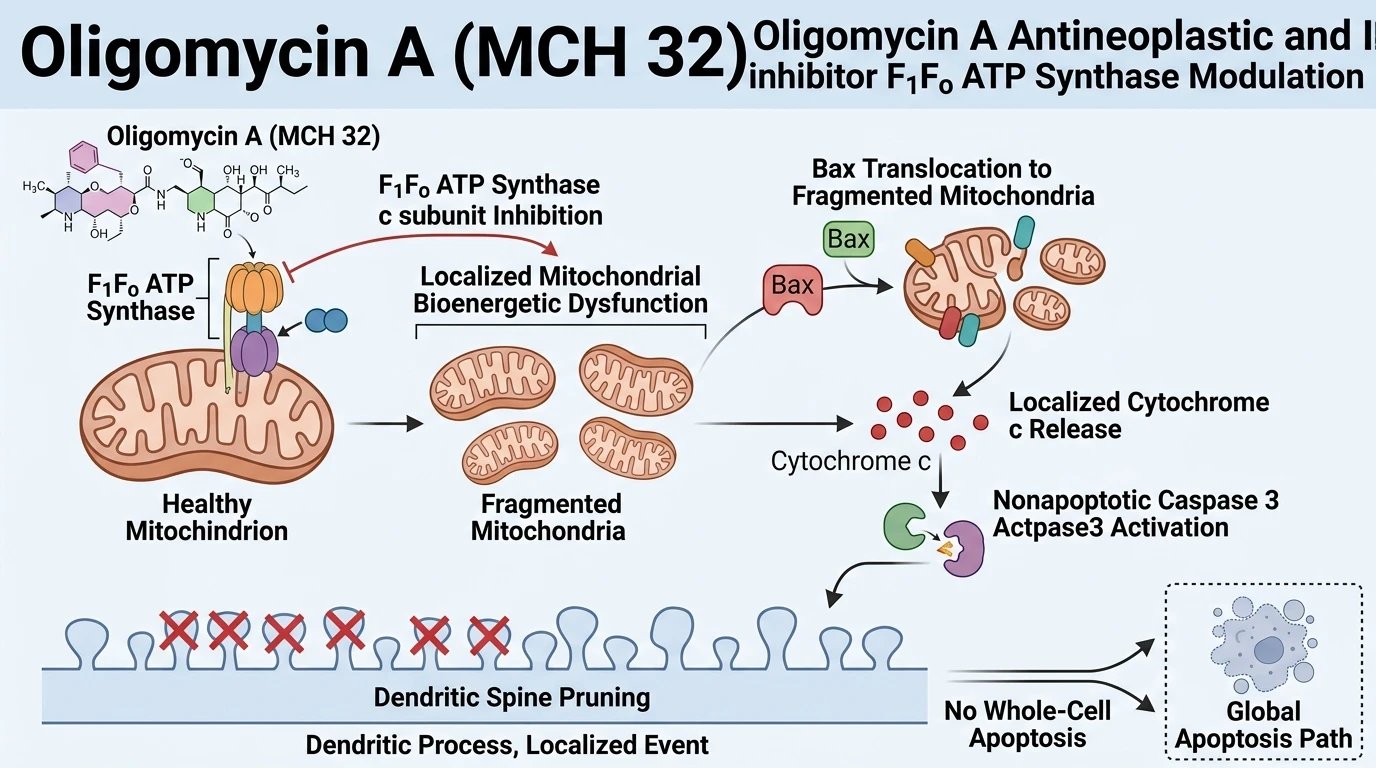
In primary hippocampal neuron models, sublethal exposure to Oligomycin A (MCH 32) at 1 µM induces rapid and severe mitochondrial dysfunction without causing immediate plasma membrane rupture or systemic necrotic cell death. Proton channel blockage causes a swift loss of mitochondrial membrane potential (ΔΨm), accompanied by a dramatic decline in basal ATP concentrations. Concurrently, the inhibition of proton flow destabilizes electron transport efficiency, generating heightened levels of mitochondrial reactive oxygen species (ROS). These metabolic disruptions directly alter organelle dynamics. Mitochondria residing in dendritic branches undergo increased fission and structural fragmentation. Furthermore, these compromised organelles exhibit reduced trafficking and diminished localization to dendritic spines, depriving localized post-synaptic structures of essential metabolic support.
Crucially, this mitochondrial distress initiates a compartmentalized, nonapoptotic biochemical cascade. Mitochondrial depolarization and elevated ROS promote the translocation of the pro-apoptotic protein Bax onto the outer mitochondrial membrane. Subsequent localized cytochrome c release occurs within dendritic segments. Rather than triggering whole-cell executioner activation, this regional cytochrome c liberation drives spatially restricted, nonapoptotic caspase 3 activation. Active caspase 3 subsequently targets localized cytoskeletal and scaffolding substrates, leading directly to the loss and morphological collapse of dendritic spines. Notably, co-treatment with the broad-spectrum caspase inhibitor Q-VD-OPh prevents spine loss and preserves dendritic architecture. However, Q-VD-OPh fails to restore mitochondrial membrane potential or ATP levels. These empirical observations establish that nonapoptotic caspase 3 activation acts as a direct, obligate downstream mediator of spine elimination triggered by F1Fo ATP synthase inhibition.
Mechanistic Synthesis of F1Fo ATP Synthase Inhibition
Copy code to add this image to your page
Oligomycin A selectively inhibits F1Fo ATP synthase by binding to the c subunit, blocking proton translocation across the inner mitochondrial membrane. In neuronal dendrites, sublethal exposure triggers bioenergetic collapse, characterized by membrane depolarization, reduced ATP production, and elevated ROS. This metabolic stress induces Bax translocation and localized cytochrome c release, activating nonapoptotic caspase 3. Localized caspase 3 cleaves structural substrates, driving dendritic spine elimination without initiating executioner caspase pathways or systemic neuronal death.
Bioenergetic Disruption Impairs Localized Dendritic Organelle Dynamics
Sublethal pharmacological inhibition using Oligomycin A (MCH 32) exposes distinct sub-cellular vulnerabilities within neuronal structures. Under primary hippocampal culture conditions, exposure to 1 µM of this compound causes immediate proton transport arrest across the F1Fo ATP synthase c subunit. Mitochondrial membrane potential collapses rapidly. Intracellular ATP pools deplete, while mitochondrial reactive oxygen species production accelerates. Furthermore, dendritic mitochondria lose their characteristic elongated morphology and undergo marked fragmentation. Quantitative imaging reveals reduced mitochondrial occupancy within dendritic spines. The localized energy crisis alters synaptic maintenance. Sub-cellular regions deprived of functional mitochondria display heightened structural instability.
Localized Nonapoptotic Caspase Activation Mediates Dendritic Spine Loss
Downstream of bioenergetic collapse, targeted outer mitochondrial membrane permeabilization occurs locally within dendrites. Bax translocates to fragmented mitochondria. Cytochrome c enters the dendritic cytosol, initiating localized nonapoptotic caspase 3 cleavage. Importantly, this compartmentalized enzymatic activity occurs without triggering global cell death or nuclear pyknosis. Pharmacological evaluation using the Oligomycin A Antineoplastic and I inhibitor paradigm demonstrates that application of the pan-caspase inhibitor Q-VD-OPh completely preserves dendritic spine density and head diameter. However, Q-VD-OPh administration does not reverse underlying mitochondrial membrane depolarization or ATP reduction. These data confirm that local caspase activity acts as an independent executioner of structural elimination following mitochondrial failure.