Camptothecin (CPT) Primed Stem Cell Vesicles Mitigate Post-Traumatic Tendon Adhesions
Copy code to add this image to your page
Post-traumatic tendon adhesion remains a major clinical challenge following musculoskeletal injury and surgical repair. Excessive proliferation of peritendinous fibroblasts and driven deposition of extracellular matrix components, predominantly stimulated by transforming growth factor-beta 1 (TGF-β1), leads to dense fibrotic scar formation. This fibrotic envelope restricts normal gliding mechanics, resulting in joint stiffness and permanent functional loss. Traditional anti-adhesive barriers physically separate tissues but fail to modulate the underlying cellular signal transduction mechanisms. Stem cell-derived extracellular vesicles offer a nanotherapeutics approach; however, unprimed naive vesicles often exhibit modest bioactivity in highly fibrotic microenvironments.
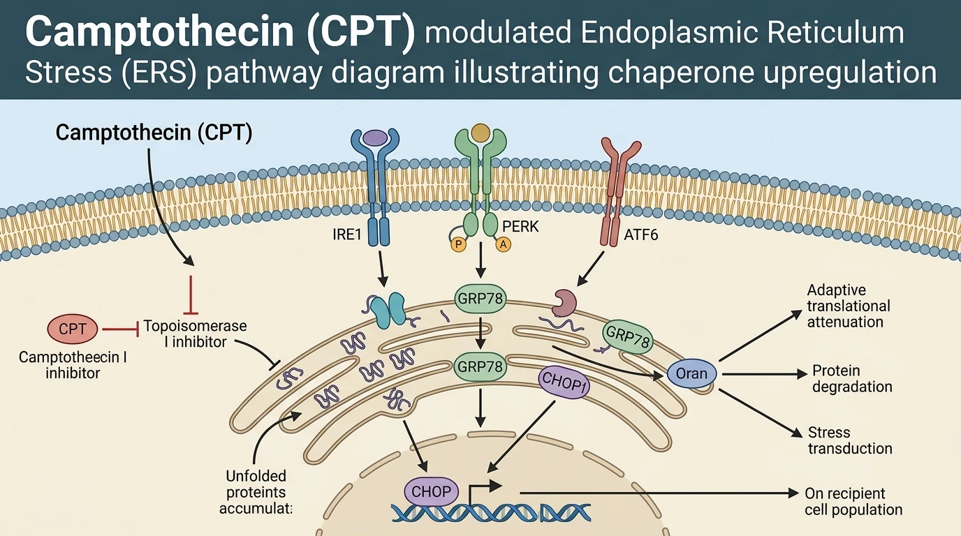
Sublethal chemical preconditioning modifies the secretome and vesicular cargo of stem cells. Applying targeted topoisomerase I inhibition via Camptothecin (CPT) derivatives—specifically hydroxycamptothecin (HCPT)—activates adaptive stress networks within human umbilical cord mesenchymal stem cells (HUMSCs). Low-dose CPT derivative priming induces controlled perturbations in protein folding homeostasis, initiating the Endoplasmic Reticulum Stress (ERS) pathway. Rather than triggering cell death cascade execution in donor cells, this sublethal stress induces a selective protein-sorting mechanism into emerging extracellular vesicles (HCPT-EVs).
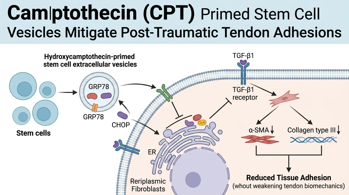
Quantitative biochemical analyses demonstrate that HCPT-EVs carry enriched concentrations of key ERS effector proteins, specifically glucose-regulated protein 78 (GRP78) and CCAAT/enhancer-binding protein homologous protein (CHOP). GRP78 functions as a major luminal chaperone, whereas CHOP acts as a transcriptional coordinator during sustained stress responses. When these enriched nanovesicles are endocytosed by recipient peritendinous fibroblasts, they transfer GRP78 and CHOP directly into the target cell cytosol. This intracellular cargo delivery re-establishes ERS signal transduction within recipient fibroblasts under TGF-β1 stimulation.
At the cellular level, activation of ERS in recipient fibroblasts fundamentally antagonizes canonical TGF-β1 signal transduction. Under tested in vitro assay conditions, treatment with HCPT-EVs markedly suppressed fibroblast proliferation rates and diminished overall cellular viability compared to unprimed EV controls. Furthermore, HCPT-EV delivery inhibited myofibroblast differentiation, characterized by a sharp reduction in alpha-smooth muscle actin (α-SMA) stress fiber assembly and decreased collagen type III (COL III) gene expression and protein deposition. By dampening the contractile phenotype of active myofibroblasts, the vesicular delivery system prevents excessive tissue contraction.
In vivo validation using a rat Achilles tendon transection and repair model confirms these cellular observations. Localized administration of HCPT-EVs within a hydrogel scaffold significantly attenuated peritendinous adhesion severity. Histological scoring revealed loose, organized connective tissue at the repair site rather than dense collagenous scar bands. Crucially, biomechanical testing indicated that biomechanical tensile strength was preserved in the regenerated tendon, showing that anti-fibrotic intervention did not compromise structural repair integrity.
Pharmacological Evaluation and Mechanistic Cargo Characterization
Copy code to add this image to your page
Priming human umbilical cord mesenchymal stem cells with hydroxycamptothecin induces targeted endoplasmic reticulum stress. This response selectively packages stress effector proteins GRP78 and CHOP into extracellular vesicles (HCPT-EVs). Upon uptake by recipient fibroblasts, HCPT-EVs trigger ERS signaling that potently counteracts TGF-β1-induced cell proliferation, viability, and myofibroblast differentiation. In rodent models, this paracrine axis reduces peritendinous fibrotic adhesions while preserving native biomechanical tensile strength.
GRP78 and CHOP Enrichment in Primed Vesicles
Sublethal exposure to Camptothecin (CPT) analogs alters stem cell protein trafficking. Endoplasmic reticulum stress sensors detect misfolded luminal proteins. Consequently, donor HUMSCs upregulate key chaperones. Proteomic evaluation confirmed elevated concentrations of GRP78 and CHOP within secreted HCPT-EVs; unprimed vesicles lacked this specific enrichment profile. Endocytosis by peritendinous fibroblasts transfers these active effectors directly into recipient cytoplasm. Western blot assays demonstrated early intracellular accumulation of both proteins within four hours post-incubation under controlled conditions.
Inhibition of Fibroblast Differentiation and Matrix Deposition
Recipient fibroblasts undergo profound phenotypic shifts following vesicle uptake. TGF-β1 exposure typically drives rapid myofibroblast conversion. HCPT-EV treatment blocked this lineage commitment. Alpha-smooth muscle actin (α-SMA) expression dropped significantly in treated cell cohorts. Simultaneously, transcript levels for collagen type III (COL III) decreased relative to control groups. Short-term viability assays showed targeted attenuation of hyperproliferative fibroblast states without inducing uncontrolled apoptosis. These cellular responses demonstrate effective interruption of the fibrotic signaling cascade in vitro.
Reduction of Peritendinous Adhesions in Rat Tendon Transection
In vivo evaluation established tangible surgical benefits. Rats subjected to complete Achilles tendon transection received local HCPT-EV applications. Macroscopic inspection at four weeks revealed minimal peritendinous tethering. Histological cross-sections confirmed organized collagen fiber alignment and reduced cellular density along repair margins. Biomechanical pull-to-failure testing demonstrated no significant drop in ultimate tensile load capacity relative to normal healing controls. Within this animal model, fibrotic adhesion prevention occurred without impairing fundamental structural tendon healing.