Emricasan (IDN-6556) Caspase inhibitor Triggers RIPK1-MLKL Necroptosis in Colorectal Cancer
Copy code to add this image to your page
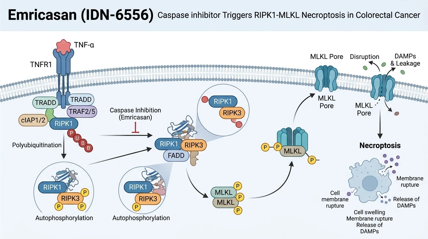
In cellular stress responses, apoptotic caspases typically clear damaged or malignant cells without inducing systemic inflammation. Pharmacological engagement of caspases by the pan-caspase inhibitor Emricasan (IDN-6556) redirects signal transduction from apoptotic machinery toward necrotic programmed cell death. In specific cellular settings, such as colorectal cancer (CRC) paradigms, blockade of catalytic caspase activity prevents caspase-8-mediated cleavage of receptor-interacting serine/threonine-protein kinase 1 (RIPK1). This pharmacological intervention preserves RIPK1 structural integrity, enabling assembly of the necrosome complex alongside RIPK3 and mixed lineage kinase domain-like protein (MLKL).
TNFAIP3-interacting protein 1 (ABIN-1, encoded by TNIP1) acts as a strict negative regulator of RIPK1-dependent signaling networks. In CRC cell lines and mouse embryonic fibroblasts, loss of ABIN-1 heightens cellular susceptibility to necroptosis-inducing combination therapies. Experimental regimens combining the Emricasan (IDN-6556) Caspase inhibitor with birinapant, 5-fluorouracil, or poly(I:C) plus the TAK1 inhibitor 5Z-7-oxozeaenol provoke pronounced necroptotic cell death. Mechanistically, these combinations stimulate autocrine secretion of tumor necrosis factor (TNF). Elevated local TNF levels engage TNF receptor 1 (TNFR1), driving assembly of downstream signaling complexes.
Under conditions of ABIN-1 deficiency, recruitment of the ubiquitin-editing enzyme A20 to the TNFR1 signaling complex is markedly diminished. Reduced A20 recruitment impairs deubiquitination, resulting in increased polyubiquitination of RIPK1. Concurrently, ABIN-1 depletion upregulates toll-like receptor 3 (TLR3) expression. Enhanced RIPK1 polyubiquitination facilitates autophosphorylation at Ser166. Active RIPK1 then recruits RIPK3, promoting cross-phosphorylation and downstream phosphorylation of MLKL at Ser358. Phosphorylated MLKL translocates to the plasma membrane, driving membrane rupture and necroptotic death.
In murine xenograft models of colorectal carcinoma, administration of IDN-6556-containing therapeutic combinations demonstrates clear tumor growth inhibition. Intratumoral biomarker analyses confirm elevated MLKL phosphorylation, verifying necroptosis execution in vivo. These observations indicate that targeting caspase activity with the Emricasan (IDN-6556) Caspase inhibitor bypasses apoptosis resistance mechanisms inherent to ABIN-1-deficient colorectal malignancies.
Pharmacological Evaluation of Caspase Inhibition and Necroptosis Activation
Copy code to add this image to your page
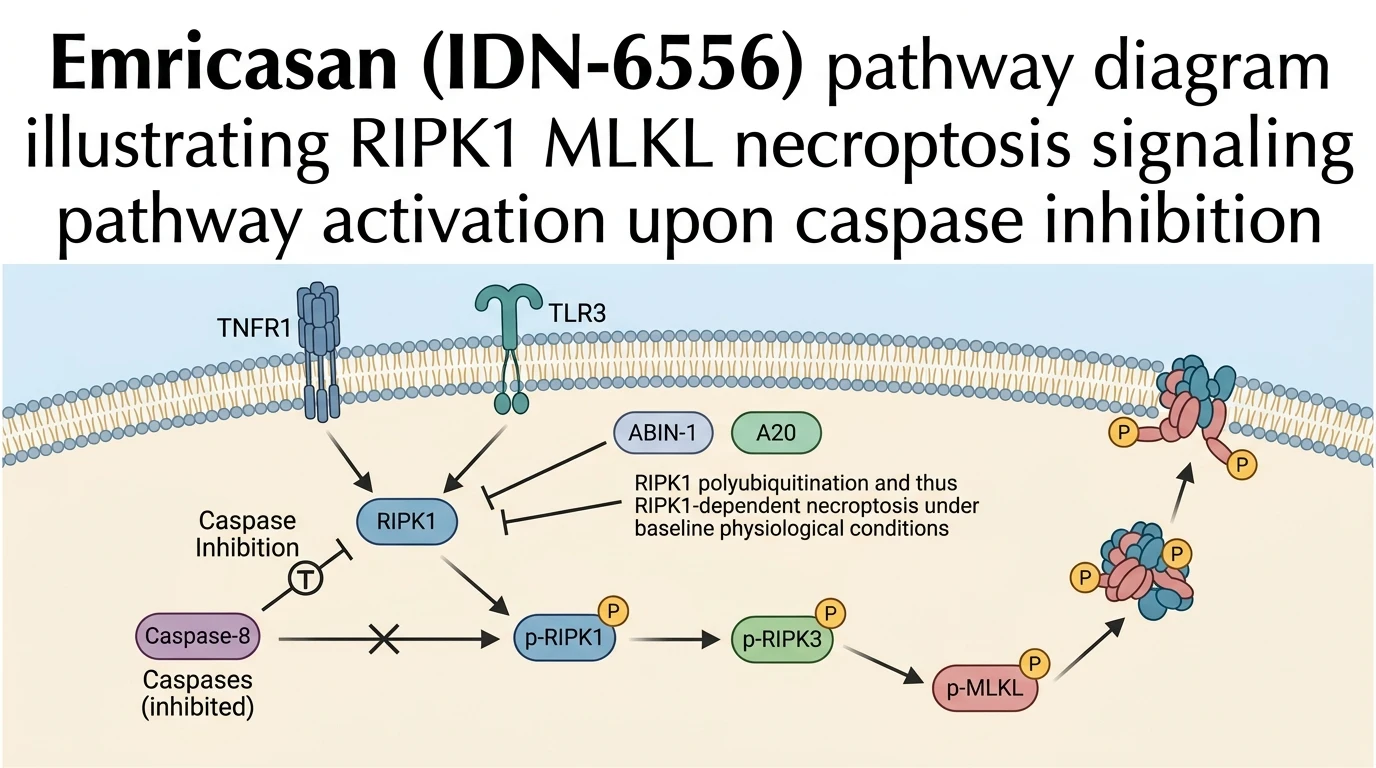
The Emricasan (IDN-6556) Caspase inhibitor blocks catalytic caspase-8 activity, preventing RIPK1 cleavage. In ABIN-1-deficient colorectal cancer cells, reduced A20 recruitment enhances RIPK1 polyubiquitination and autophosphorylation. Uncleaved RIPK1 forms a functional necrosome with RIPK3, phosphorylating MLKL. Phosphorylated MLKL oligomerizes and permeabilizes the plasma membrane, executing necroptosis.
ABIN-1 Loss Enhances RIPK1 Polyubiquitination and Necrosome Assembly
ABIN-1 restricts RIPK1 activation by facilitating A20 recruitment to TNFR1 signaling complexes. In ABIN-1-deficient colorectal cells, A20 engagement drops significantly under tested in vitro conditions. This loss of ubiquitin editing promotes persistent RIPK1 polyubiquitination. RIPK1 autophosphorylation ensues within hours of treatment. The Emricasan (IDN-6556) Caspase inhibitor prevents caspase-8 from cleaving phosphorylated RIPK1. Uncleaved RIPK1 accumulates rapidly inside the cell. Phosphorylated RIPK1 binds RIPK3 with high affinity. This interaction triggers downstream MLKL phosphorylation at Ser358. Membrane insertion of MLKL oligomers causes immediate cell swelling and plasma membrane lysis.
Synergistic Regimens Drive Autocrine TNF Secretion and Cell Death
Combining pan-caspase inhibition with targeted agents amplifies necroptotic signaling. In cultured colorectal cancer models, co-administration of the Emricasan (IDN-6556) Caspase inhibitor with birinapant or 5-fluorouracil accelerates autocrine TNF secretion. Secreted TNF acts in an autocrine loop to re-engage surface TNFR1. Concurrently, TLR3 expression rises in ABIN-1-depleted settings. Dual engagement of TNFR1 and TLR3 intensifies downstream kinase activity. Intracellular ATP levels decline precipitously following MLKL phosphorylation. In mouse xenograft models, this combination regimen significantly restricts tumor expansion compared to single-agent controls. Pharmacodynamic assays confirm robust MLKL activation across xenograft tissue sections.