Crenigacestat (LY3039478) Crenigacestat inhibitor Microenvironment Remodeling in Cholangiocarcinoma
Copy code to add this image to your page
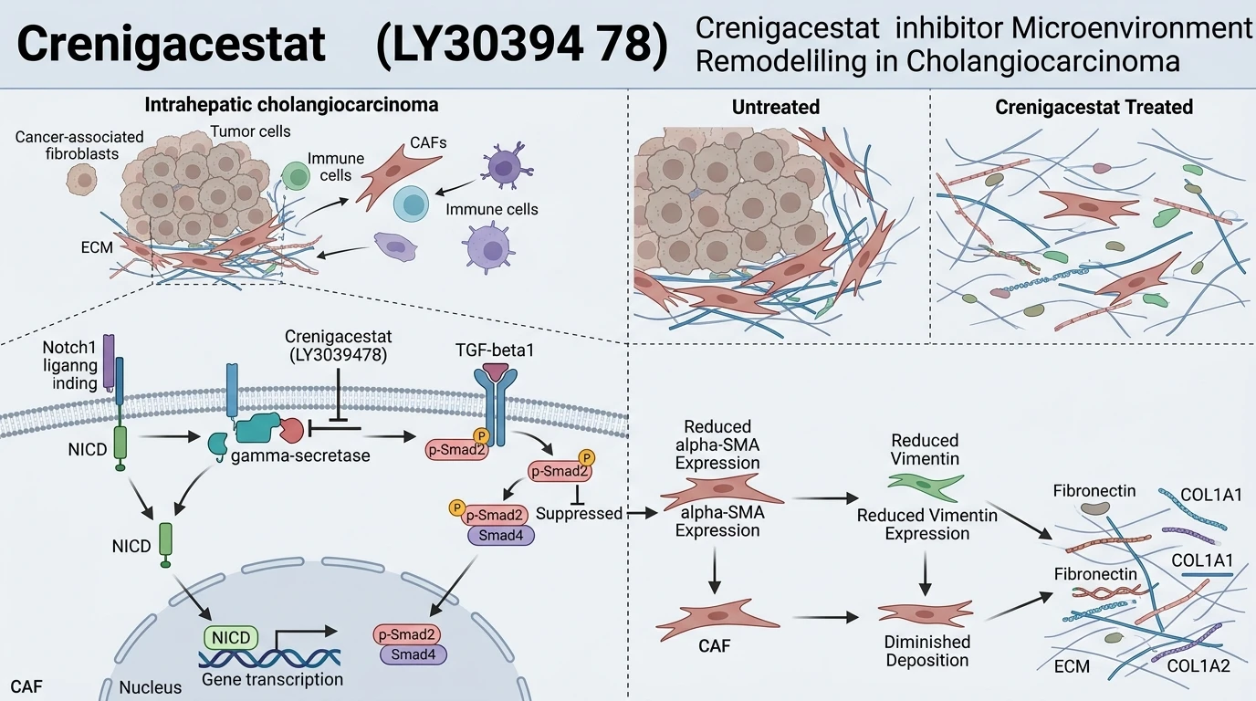
Intrahepatic cholangiocarcinoma (iCCA) exhibits an extensive desmoplastic stroma rich in activated cancer-associated fibroblasts (CAFs). This dense fibrotic extracellular matrix (ECM) poses a substantial physical barrier to drug delivery and supports tumor progression. Notch signaling plays a direct regulatory role in initiating and sustaining stromal activation within the liver microenvironment. Specifically, the processing of Notch receptors by the multi-subunit protease γ-secretase liberates the intracellular domain to drive downstream transcriptional networks. Crenigacestat (LY3039478) functions as a potent, selective oral Notch1/γ-secretase inhibitor. Through targeted binding within the catalytic complex of γ-secretase, this compound effectively halts intramembrane proteolysis.
The pharmacological activity of the preclinical evaluation of γ-secretase inhibition demonstrates high selectivity across Notch receptor isoforms. In cellular assays, exposure to Crenigacestat (LY3039478) leads to a dose-dependent reduction of cleavage products. Consequently, transcription of canonical downstream effectors decreases markedly. Beyond direct cell-intrinsic antiproliferative activity, targeting Notch signaling alters paracrine signaling loops operating between tumor cells and surrounding stromal elements. In experimental models of liver injury and malignancy, aberrant Notch signaling correlates directly with elevated transforming growth factor-beta 1 (TGF-β1) expression. Blockade of Notch processing interrupts this paracrine crosstalk.
Experimental characterization of Crenigacestat (LY3039478) highlights its capacity to attenuate peritumoral fibrosis in organoid and xenograft settings. Within intrahepatic cholangiocarcinoma tissues, the desmoplastic reaction relies heavily on continuous TGF-β1 activity. Crenigacestat treatment reduces TGF-β1 transcript levels, subsequently downregulating phosphorylation of canonical Smad2 effectors. As phosphorylated Smad2 levels decline, downstream activation pathways in hepatic stellate cells and resident fibroblasts are blunted. This signaling suppression restricts phenotypic transition into contractile, matrix-secreting CAFs.
At the cellular level, deactivated CAFs exhibit marked alterations in structural and secretory profiles. Expression of key mesenchymal markers, including alpha-smooth muscle actin (α-SMA) and Vimentin, drops substantially following treatment under controlled in vitro culture conditions. Concurrently, production of major extracellular matrix proteins decreases. Specifically, transcription and deposition of Fibronectin, collagen type I alpha 1 (COL1A1), and collagen type I alpha 2 (COL1A2) decline in a concentration-dependent manner. This loss of structural matrix components reduces tissue stiffness and alters interstitial pressure dynamics within the tumor mass.
In vivo observations in murine orthotopic and xenograft models of iCCA validate these biochemical mechanics. Administration of Crenigacestat (LY3039478) yields a noticeable reduction in peri-tumoral desmoplasia. Fibrotic tract density diminishes upon histology analysis. Furthermore, remodeling of the dense stromal matrix enhances intratumoral perfusion. When combined with standard chemotherapeutic regimens, such as Gemcitabine, Crenigacestat produces pronounced synergistic tumor growth inhibition. The reduction in extracellular matrix density facilitates improved drug penetration into previously hypoperfused tumor cores. These empirical findings illustrate that targeted Notch pathway inhibition acts as a dual mechanism agent—simultaneously targeting malignant epithelial cell signaling and modulating supportive microenvironmental stroma.
Pharmacological Evaluation and Microenvironmental Remodeling Mechanics
Copy code to add this image to your page
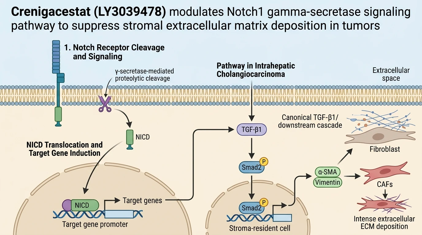
Crenigacestat (LY3039478) is a selective Notch1/γ-secretase inhibitor that blocks Notch processing and suppresses downstream TGF-β1/Smad2 signaling in intrahepatic cholangiocarcinoma. This pathway blockade deactivates cancer-associated fibroblasts, suppressing α-SMA and Vimentin expression while markedly decreasing extracellular matrix proteins, including Fibronectin, COL1A1, and COL1A2. By reducing peritumoral fibrosis and microenvironmental desmoplasia, Crenigacestat enhances tissue perfusion and acts synergistically with Gemcitabine.
Notch Blockade Suppresses Stromal TGF-β1/Smad2 Cascade
Targeting γ-secretase cleavage disrupts pivotal paracrine signaling networks within the tumor microenvironment. Within tested intrahepatic cholangiocarcinoma models, the Crenigacestat inhibitor downregulates TGF-β1 ligand secretion. Smad2 phosphorylation drops rapidly in adjacent stromal cells. This inhibition breaks the feed-forward loop sustaining chronic fibroblast recruitment. Consequently, canonical Smad2 nuclear translocation decreases significantly. Cellular transcription of profibrotic genes ceases. The observed response remains consistent across multiple patient-derived cell models.
Fibroblast Deactivation Reduces Extracellular Matrix Deposition
Activated cancer-associated fibroblasts drive the mechanical stiffness characteristic of desmoplastic tumors. Upon exposure to Crenigacestat (LY3039478), stromal fibroblasts undergo phenotypic deactivation. Expression of contractile markers α-SMA and Vimentin decreases markedly. In parallel, secretome analysis reveals diminished accumulation of key extracellular matrix scaffolding proteins, specifically Fibronectin, COL1A1, and COL1A2. The surrounding physical matrix becomes measurably less dense. Interstitial fluid pressure declines within primary tumor tissue.
Combinatorial Synergism with Gemcitabine in Vivo
Microenvironmental remodeling directly enhances the delivery of co-administered cytotoxic agents. In murine xenograft models of intrahepatic cholangiocarcinoma, monotherapy with Gemcitabine often yields constrained responses due to stromal exclusion. Co-administration of the Crenigacestat inhibitor significantly increases intratumoral accumulation of Gemcitabine active metabolites. Tumor volume regression exceeds effects achieved by either single agent alone. Survival metrics improve in treated animal cohorts. These empirical observations highlight microenvironmental normalization as a practical strategy to overcome chemoresistance.